Steps:
1. Cross the nicols, and find a mineral with low birefringence.
2. Focus the mineral using higher objective (40X).
3. Insert condensing lens (For conoscopic view)
4. Insert Bertrand lens or remove ocular.
click here to see a uniaxial figure
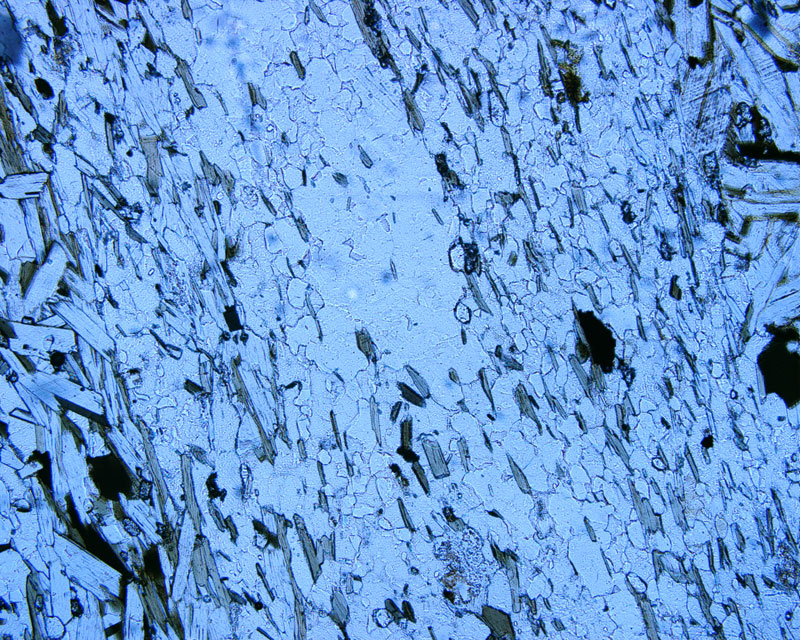
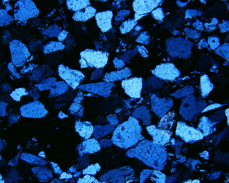
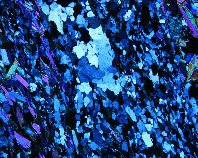
I. Following the above steps obtain an interference figures from BH-250-35 (quartz (1,2,3,4,5,6) ) perpendicular to C slides and Calcite BH-250-36 perpendicular to C slides. (Take a picture of your figures and turn in to you TA or instructor.)
Note that there are two components to the uniaxial interference figure.
They are the bands of extinction, the dark zones, called isogyres, and bands
of equal colors, termed isochromatic curves. Now rotate the stage. If the crystal
section is oriented perfectly perpendicular to the optic axis, the figure will
remain stationary. If the crystal is oriented at some angle to the optic axis,
the interference figure will rotate with the stage.
a. Does your figure rotate as you rotate the stage?
b. What interference color do you observe when viewing the calcite in orthoscopic
configuration?
II. Sketch the optic axis figure you see in the calcite section. Be sure to
show the number of alternating dark and light colored isochromatic curves you
see and the orientation of the isogyres.
Click
here to print entire optics lab 7